


CASE 2
Socket grafting ("socket fill”) in the maxilla and dental implantsPatient: female, 61 years
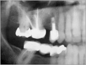
2.1
2.2
2.3
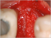
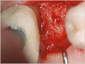
2.2 Intraoperative situation two weeks after tooth extraction and after socket grafting with phycogenic apatite.
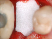
2.3 Covering with collagen membrane
2.4
2.5
2.6
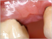
2.5 Primary wound healing with a perfect mucosa level which means perfect ridge preservation and enables insertion of a dental implant, which was inserted five months later.
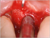
2.6 After opening the mucosa there are only residues of the apatite granules to observe which will not be removed because later they contribute to the stabilisation of the periimplantary mucosa.
2.7
2.8
2.9
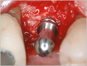
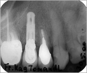
2.8 Situation after implantation following drill hole widening with condensors.
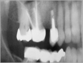
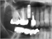
2.9 Panorex after 6 months just before reopening the implant.
2.10
2.11
2.12
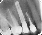
2.11 Dental X-ray after six years.
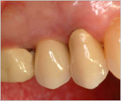
2.12 Periimplant situation after six years.