CASE 6

Sinus elevation and augmentation with phycogenic apatite and dental implants
Patient: female, 56 years
Augmentation of phycogenic apatite in combination with collector bone, PRP and thrombin.

AlgOss - clinical use
 6.1
AlgOss - clinical use
 6.2
 
6.1 Panorex immediately after bilateral sinus elevation and sinus grafting with phycogenic apatite, collector bone, PRP and thrombin on both sides.

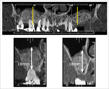
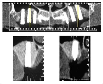
6.2 Panorex view of a reformatted Dental CT immediately after surgery. Below are the reformatted orthoradial slices 13 and 43 with about 18 mm vertical height of the applied augmentation material. Short after the operation the phycogenic material is only little radioopaque. When using thrombin we always observe bubbles in the augmentation material.

AlgOss - clinical use
 6.3
AlgOss - clinical use
 6.4
 
6.3 Panorex view of a reformatted Dental CT 6 months after surgery. Below are the reformatted orthoradial slices 10 and 38 with about 16 and 18 mm vertical height of the applied augmentation material. During the 6 months healing time the augmentation material became calcified and is therefore more radioopaque. The bubbles have vanished and the material impresses as solid newly formed bone.

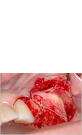
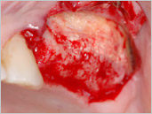
6.4 Reopening to remove the non resorbable e-PTFE membrane after sinus elevation and sinus grafting. Non resorbable membrane still in situ.

AlgOss - clinical use
 6.5
AlgOss - clinical use
 6.6
AlgOss - clinical use
 6.7
 
6.5 After removal of the non resorbable e-PTFE membrane the completely integrated and into newly formed bone transformed phycogenic apatite is visible. Fresh extraction socket of tooth Nr. 12.

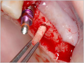
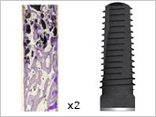
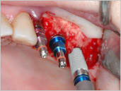
6.6 Behind the immediate inserted implants in region tooth Nr. 12, harvesting of a core sample for histology before insertion of the implant in region Nr. 13.

6.7 Removing of the core sample for histology.

AlgOss - clinical use
 6.8
AlgOss - clinical use
 6.9
AlgOss - clinical use
 6.10
 
6.8 Hard tissue slides of the core sample in two times magnification with sufficient newly formed bone in the surroundings of the phycogenic augmentation material after six months healing time. Into the trephine hole the implant gets inserted.

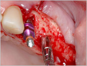
6.9 Situation after insertion of an immediate implant in region Nr. 12, insertion of an implant in region Nr. 13 and before inserting the implant into region Nr. 14.

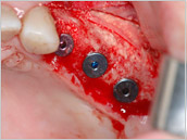
6.10 Situation after applying the cover screws and filling the gap around the implant in region 12 with the phycogenic augmentation material.

AlgOss - clinical use
 6.11
AlgOss - clinical use
 6.12
 
6.11 Primary suture closure with non resorbable suture material after palatinal incision.

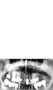
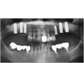
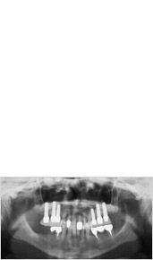
6.12 Panorex after insertion of 6 implants.

 
AlgOss - clinical use
 6.13
AlgOss - clinical use
 6.14
 
6.13 Reformatted panorex view of the dental CT after implant insertion. Below are the reformatted orthoradial slices 14 with a 4.3 mm thick and 16 mm long implant and 46 with a 5 mm thick and 16 mm long implant visible.

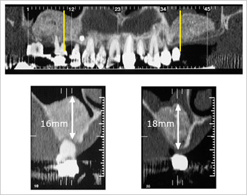
6.14 Panorex 3.5 years after sinus elevation and sinus grafting.

AlgOss - clinical use
 6.15
 
6.15 Situation 3.5 years after sinus elevation and sinus grafting.