


CASE 5
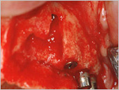
Augmentation of large defect in the maxilla and implantationPatient: female, 73 years
5.1
5.2

5.3
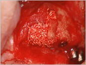
5.2 Augmentation with a small monocortical onlay graft from the tuberosita and filling with phycogenic apatite.
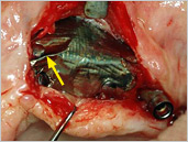
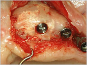
5.3 Onlay and augmentation material covered and stabilised with a titanium membrane fixed with 3 titanium pins. Please note the wrinkle in the titanium membrane (arrow).
5.4
5.5
5.6
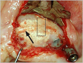
5.5 Filled defect with newly formed bone. The osseointegrated but not completely resorbed phycogenic material shines through the newly formed bone. The gap under the wrinkle of the titanium membrane has been filled with newly formed bone (arrow).
5.6 After implant insertion.
5.7
5.8
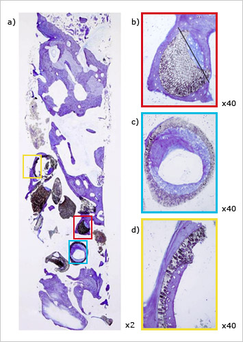
a) Two times magnification of the core specimen. Below = alveolar ridge, top = apical region.
b) Red box: 40 times magnification.
c) Blue box: Transverse section of b) (black line) also in 40 times magnification.
d) Yellow box: 40 times magnification from a). Excellent new bone formation around and in between the granule with little signs of beginning resorption.
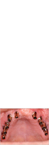
5.8 Telescopic crowns 9 years later.